Thomae Sourlingas, Senior Researcher B’ Grade
Kalliope Sekeri, Emeritus, A’ Grade
Marios Xydous, Postdoctoral Collaborator
Elena Kostopoulou, Postgraduate Student – MSc obtained in 2012
Paraskevi Salpea, Postgraduate Student – PhD obtained in 2011
Yannis Ninios, Postgraduate Student – PhD obtained in 2009
Research Interests
The research interests of our group are focused on the study of the role of
1) histone proteins and their subtypes,
2) histone epigenetic post translational modifications and
3) histone deacetylase inhibitors in chromatin function during various biological processes.
-
Ageing / Senescence: Investigation of the changes in the expression profiles of the H1 DNA linker histone subtypes and in histone post translational modifications (phosphorylation, acetylation and methylation) during ageing in human leucocytes, cell strains and cell lines. We are also studying the changes that these histone epigenetic modifications bring about in the expression profiles of age-dependent genes.
-
Apoptosis: Study of the interactions and functional role of specific H1 DNA linker histone subtypes with the DNA fragmentation factor, DFF40, an endonuclease that cleaves DNA during the last stages of apoptosis. This line of research is being carried out in human leukemic cell lines after the induction of apoptosis with histone deacetylase inhibitors. The effects of histone deacetylase inhibitors in the acetylation of histones and non histone target molecules are also being studied. The aim of these studies is to find molecules and/or factors which may have a functionally active involvement during the course of apoptosis.
-
Mammalian Biological Clock: Investigation of the role of chromatin conformational changes that are brought about by histone post translational epigenetic modifications, such as histone acetylation and methylation, in the regulation of circadian gene expression and in mammalian clock function.
-
Psychiatric Disorders: Analysis of how changes in histone epigenetic modifications and in the expression profiles of the H1 DNA linker histone subtypes affect chromatin remodelling (conformational reorganization) events in human peripheral blood leucocytes from individuals with psychiatric disorders.
CURRENT RESEARCH PROJECTS
Ageing / Senescence
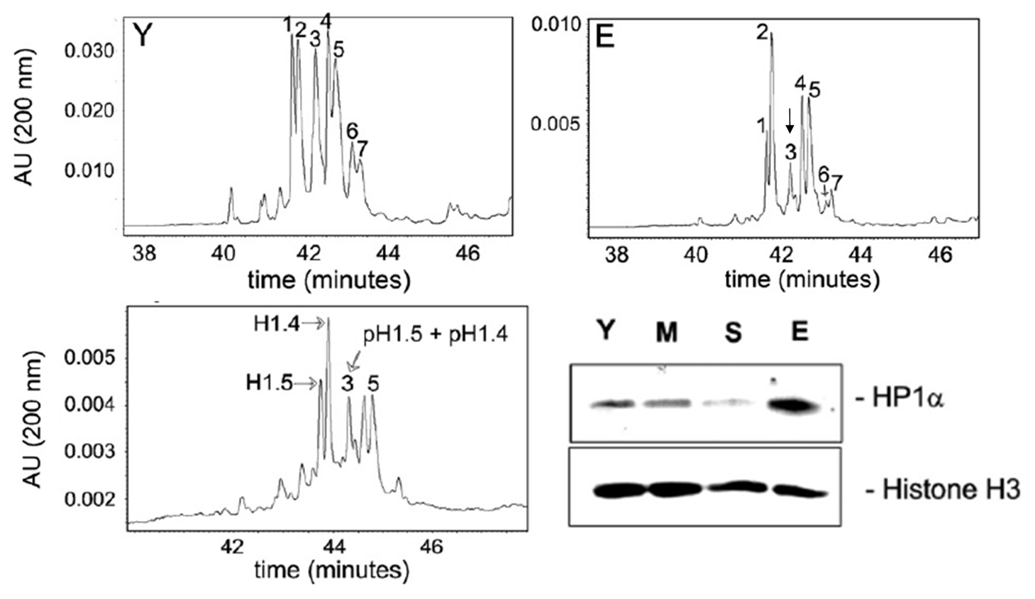
The dephosphorylation of the histone linker H1 subtypes, H1.4 and H1.5, is associated with an increase in heterochromatin formation, i.e., inactive chromatin as a function of increasing donor age.

Figure 1: Separation of the H1 subtypes by Capillary Zone Electrophoresis (CZE), where a decrease in peak no. 3 was found in the resolved H1 histone fraction in lymphocytes from elderly donors with respect to that of young donors. The peaks were identified and peak no. 3 was shown to correspond to the phosphorylated isoforms of H1.4 and H1.5 (pH1.4, pH1.5). Concomitantly, western blot analysis showed that there was an increase in the heterochromatin protein, HP1α, in the lymphocytes of the same elderly donors. Y = young, M = mid-aged, S = senior, E = elderly. Histone H3, reference loading protein control (Happel, N., Doenecke, D., Sekeri-Pataryas, K.E. abd T.G. Sourlingas, Exp. Gerontol., 2008).
This line of work associated for the first time the dephosphorylation of two H1 subtypes with an increase in heterochromatin formation during ageing and senescence in human peripheral blood lymphocytes. Given the fact that phoshorylation – dephosphorylation, as well as the presence of specific H1 histone subtypes play a significant role in chromatin conformational changes during various biological processes, including ageing (as indicated by the findings of the present investigation), it may be concluded that heterochromatinization is a necessary process during lymphocyte ageing which leads to a gradual shutting down of non-essential cellular processes to the bare minimum, for cellular economization and survival by maintenance of the senescent phenotype. Future work for this research project will be aimed at investigating whether the post translational phosphorylation profile, as well as that of other histone epigenetic modifications play a similar role in heterochromatin formation during the ageing process of other cellular / tissue systems.
Apoptosis
(1) Results from our group showed that the DNA fragmentation factor (DFF40), which is activated during the final stages of apoptosis is located on the chromatin fraction, not only in its active form after the caspase-dependent proteolysis of DFF45 after the apoptotic stimulus, but also in its inactive form as a heterodimer with its chaperone, DFF45. It was already known that DFF40 interacts with H1 linker histone, which recruits DFF40 to the linker DNA region, resulting in internucleosomal DNA fragmentation in in vitro cell-free systems. But it was not known whether DFF40 may interact in a preferential manner with specific H1 histone subtypes. Results from our lab showed that DFF40 associates with all the H1 subtypes, but preferentially interacts with specific subtypes, i.e., H1.5 and H1.0, while its association with H1.3 was found to be decreased.

Figure 2: Relative chromatin composition of the H1 subtypes, H1.1, H1.3, H1.5 and H1.0, (A) and relative quantification of the H1 subtypes after co-immunoprecipitation (co-IP) with DFF40 of the chromatin fraction of the NB4 leukemic cell line (B). The results are expressed as the ratio of each H1 subtype to the total H1 fraction. TSA, trichostatin A (histone deacetylase inhibitor), apoptotic stimulus (Ninios, Y.P., Sekeri-Pataryas, K.E. and T.G. Sourlingas, Apoptosis, 2010).
These results are of importance since, for the first time, the association of DFF40 with individual H1 subtypes and under a specific apoptotic external stimulus have been analyzed in an in vivo cellular context (the promyelocytic leukemic cell line, NB4). It remains to be seen whether this is specific to the cancer cell type and/or whether the specific apoptotic agent used is a determining factor in the degree of the association of DFF40 with specific H1 subtypes.
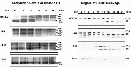
(2) In this project we used the histone deacetylase inhibitor (HDACI), trichostatin A, which causes the accumulation of acetylated histones, as well as that of certain non-histone proteins, such as α-tubulin, as the apoptotic stimulus in four leukemic cell lines and found that the degree of acetylation reflects the level of apoptosis induced in each cell line. These findings are of special significance since, though it has already been reported that apoptosis induced by histone deacetylase inhibitors may be correlated with increased histone acetylation and hyperacetylation in many cell systems, the opposite has not been adequately investigated, i.e., whether HDACIs induce apoptosis in a cell line that is resistant to the accumulation of acetylated histones in the presence of these agents. We found that the K562 leukemic cell line, which is resistant to the induction of apoptosis by trichostatin A does not show changes in the acetylation levels neither of histones nor α-tubulin. Our findings indicate that the accumulation of acetylated substrates in the continued presence of TSA seems to be a necessary factor for the initiation and completion of the apoptotic process. Given the wide use of HDACIs in anticancer regimens, alone or in conjunction with other anticancer agents, our results indicate that the evaluation of the acetylation levels of target proteins by HDACIs may possibly have the potential of being used as additional indicators of the responsiveness or sensitivity of different cancer cell types to this continuously growing class of anticancer agents.

Figure 3: Levels of histone H4 acetylation after the electrophoretic resolution of the H4 isoforms and the degree of apoptosis by western blot analysis for the detection of the levels of PARP cleavage. TSA, trichostatin A. Η4.0, Η4.1, Η4.2, Η4.3 and H4.4. non-, mono-, di-, tri- and tetra-acetylated histone H4 isoforms, respectively (Ninios, Y.P., Sekeri-Pataryas, K.E. and T.G. Sourlingas, Leuk. Res., 2009).
TSA is a highly potent and specific HDAC inhibitor, which has been and will continue to be a valuable tool for the in vitro study of the effects of histone deacetylase inhibitors. However it is not used clinically. Future work for this project will be aimed at investigating other histone deacetylase inhibitors which are in a pre-clinical phase and/or in clinical use using a wider range of cancer cell types / tissues in order to ascertain whether protein acetylation levels may constitute additional reliable biomarkers for the effectiveness of this class of apoptotic agents in anticancer schemes.
The Mammalian Biological Clock
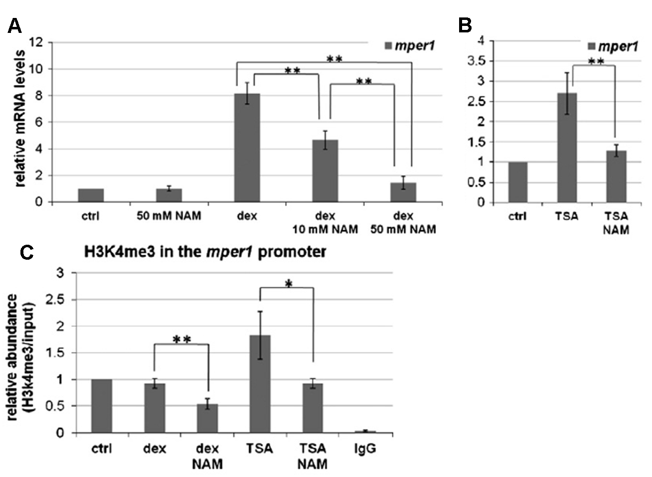
We examined the effects that two histone deacetylase inhibitors (HDACs), nicotinamide and trichostatin A (TSA), have on circadian clock function in NIH3T3 cell cultures (immortalized mouse fibroblasts). Real-time PCR showed that TSA increases the mRNA levels of the early response clock gene per1 (period1) and that nicotinamide inhibits this TSA-induced effect. Moreover, nicotinamide blocks the acute response of per1 induced by dexamethasone. This response is necessary for the synchronization of the circadian clock in cell cultures. In both cases (TSA, dexamethasone), nicotinamide’s inhibitory effects are not directly associated with its ability to inhibit class III histone deacetylases (sirtuins), due to the fact that use of another alternate inhibitor of this class of deacetylases does not lead to suppression of per1 induction. ChIP experiments showed that inhibition of the acute response, as well as the increase in per1 mRNA levels induced by TSA, is associated with a decrease in the lysine 4 trimethylation levels of histone H3 (H3K4me3) in the promoter region of this gene.

These results reveal the existence of a novel, as yet to be reported, mechanism with which clock function is regulated. Future work for this project will be aimed at unraveling whether the mechanisms by which nicotinamide decreases H3K4me3 levels are associated with changes in methylase or demethylase activity. Aside from addition of new data in the field of gene regulatory mechanisms, the elucidation of the molecular basis by which nicotinamide affects the trimethylation levels of H3K4 may prove to be of additional significance to human and livestock nutrition since nicotinamide is widely used as a nutritional supplement (vitamin B3 / niacin).


